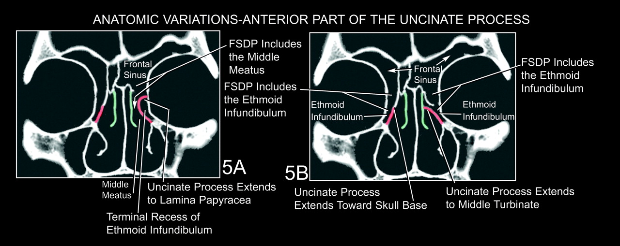
Fig 5.

Anatomic variations in the anterior uncinate process.
A and B, Coronal CT sections modified from the skull illustrated in Figs 3 and 4. Coronal CT sections display the three major variations in the superior attachment of the uncinate process (7): extension laterally to join the lamina papyracea (A, left), extension superiorly to join the skull base (A and B, right), and extension medially to join the lateral surface of the middle turbinate (B, left). Because of these variations in the uncinate process, the frontal sinus drainage on the left side of panel A passes through the superior compartment directly into the middle meatus, not the ethmoid infundibulum. In contrast, the frontal sinus drainage on the right side of panel A and both sides of panel B passes through the superior compartment into the ethmoid infundibulum before entering the middle meatus. The ethmoid infundibulum forms the inferior compartment of the FSDP in these cases. In A, the attachment of the left uncinate process with the lamina papyracea forms a terminal recess of the ethmoid infundibulum.


Related Articles
Cited By...
- No citing articles found.