Abstract
SUMMARY: Various facial reanimation procedures can be performed for treating patients with chronic facial nerve paralysis. The radiologic imaging features of static and dynamic techniques are reviewed in this article with clinical correlation, including brow lift, eyelid weights and springs, gracilis free flaps, fascia lata grafts, temporalis flaps, and Gore-Tex suspension slings. Although the anatomic alterations resulting from facial reanimation surgery may not necessarily be the focus of the imaging examination, it is important to recognize such changes and be familiar with MR imaging compatibility of the associated implanted materials. Furthermore, imaging is sometimes used to specifically evaluate the postoperative results, such as vessel patency following free gracilis transfer.
Chronic facial nerve paralysis can result in significant morbidity, including brow ptosis, lagophthalmos, ectropion, exposure keratopathy, nasal alar collapse, effacement of the nasolabial fold, ptosis of the oral commissure, and drooling. Static and dynamic facial reanimation surgical techniques are available to prevent and treat these complications. Static facial nerve rehabilitation procedures include brow lift, eyelid weight implantation, lower eyelid canthoplasty and tightening, fascia lata and alloplastic slings, and cheiloplasty. Dynamic facial reanimation procedures include nerve transfer and grafting, eyelid springs, free muscle transfer, regional muscle transfer, and lengthening myoplasty. The anatomic changes brought about by these procedures can be delineated by using conventional radiologic imaging modalities, including radiographs, CT, sonography, and MR imaging. Furthermore, in certain instances, imaging may be requested specifically to evaluate the results of facial reanimation surgery, such as interrogating the patency of the vascular pedicle following gracilis muscle transfer. The imaging findings after selected static and dynamic facial reanimation surgeries are described and depicted in the following sections.
Brow Lift
Brow ptosis can be addressed by performing a brow lift. Several methods exist to elevate the brow, one of which consists of implanting a fixation device into the frontal calvaria to which a permanent suture is secured (Fig 1).1 Brow lift can be performed in conjunction with ablation of the brow depressor muscles and blepharoplasty. Numerous fixation devices can be used, including pins, screws, tacks, K-wires, and tissue adhesives. Metallic fixation devices can be depicted on CT and should not be mistaken for unintended foreign bodies (Fig 2).
Brow lift. Intraoperative photographs show screws in the frontal bone used to secure sutures tunneled under the subcutaneous tissues toward the brow.
Brow lift. Axial CT image shows a metallic left frontal bone pin (arrow) used for suture fixation.
Eyelid Weights
Implantation of gold and platinum eyelid weights into the upper eyelid is a static reanimation procedure for treating lagophthalmos.2 Although gold weights are more commonly used, platinum weights have a thinner profile and therefore offer an increased aesthetic benefit.3 Available eyelid implant designs include curved metal sheets with holes to permit anchoring with a suture (Fig 3) and flexible metal chains. In-growth of fibrous tissue through the holes also helps secure the weight in position. Eyelid weights are secured to the superficial aspect of the upper eyelid tarsal plate. The eyelid weights generally produce considerable streak artifacts on CT, which can obscure surrounding structures (Fig 4). Platinum and gold eyelid weights are considered MR imaging–compatible up to 7T4,5 but may cause local field inhomogeneity (Fig 5). Complications related to eyelid weight implantation include suboptimal eyelid contour, infection, allergic reaction, migration, and extrusion.6
Eyelid weight. Frontal radiograph shows a gold implant containing 3 drill holes at the level of the left upper eyelid.
Eyelid weight. Axial CT image shows extensive streak artifacts related to the left eyelid weight, which obscures surrounding structures.
Eyelid weight. Axial post-contrast fat-suppressed T1-weighted image shows field inhomogeneity associated with the left eyelid weight (arrow).
Eyelid Springs
Eyelid springs are used to augment eyelid closure in patients with eyelid paralysis.7,8 The spring has the ability to achieve complete eye closure in the supine position.9 The device is implanted via orbitotomy and consists of a palpebral branch and an orbital branch connected by a spring mechanism at the fulcrum. The springs are generally composed of stainless steel and are MR imaging–compatible up to at least 1.5T.10 The positioning and function of the device can be evaluated on radiographs obtained in the open and closed lid positions, whereby the palpebral branch is expected to descend with lid closure (Fig 6). Potential complications of eyelid springs include dislocation, metal fatigue resulting in failure, and exposure.
Eyelid spring. Frontal (A) and lateral (B) radiographs show that the inferior limb of the spring is positioned in the superior eyelid (arrows), while the superior limb is positioned along the orbital rim.
Gracilis Free Flap
Free gracilis transfer is an effective method of smile rehabilitation for facial paralysis in selected patients (Fig 7).11⇓⇓–14 The gracilis muscle is harvested from the thigh with its neurovascular supply (Fig 8). The graft is then inset in the plane deep to the superficial musculoaponeurotic system and extends from the zygomatic arch to the modiolus of the oral commissure (Fig 9). The vascular pedicle of the graft is typically anastomosed to the facial artery and vein, and the obturator nerve can be anastomosed to a cross–face nerve graft and/or the masseteric branch of the trigeminal nerve.15,16 Vascular ring coupler devices are often used for the venous anastomosis.17 Depending on the particular type, the ring coupler may appear as a hyperattenuated circular structure overlying the angle of the mandible (Fig 10). Postoperative flap monitoring by physical examination alone is challenging and is often supplemented with use of a hand-held Doppler probe. Color Doppler sonography is an effective and noninvasive tool for evaluating arterial and venous flow through the pedicle of the buried free flap, whereby a sharp systolic upstroke should be evident in the artery and continuous flow should be observed in the vein (Fig 11). The examination can potentially avoid wound exploration to verify appropriate muscle perfusion and is typically performed on the first postoperative day.18 The arterial waveform of the graft should normally demonstrate a sharp systolic upstroke, while the vein may normally exhibit a continuous waveform and should be compressible, except at the site of the ring connector device. A good functional outcome correlates with normal muscle structure of the free flap depicted on MR imaging.19 Imaging may also be useful for measuring the graft thickness,20 which potentially relates to function.
Gracilis free flap. Clinical photographs before (A) and after (B) gracilis free flap reanimation show marked restoration of the patient's smile.
Gracilis free flap. Intraoperative photograph shows the harvested flap (asterisk) with an attached vascular pedicle (arrow) and nerve (arrowhead).
Gracilis free flap. Axial (A) and coronal (B) postcontrast T1-weighted MR images show a healthy gracilis flap (arrows), which extends from the zygomatic arch to the oral commissure following total left parotidectomy, with facial nerve sacrifice for resection of mucoepidermoid carcinoma.
Gracilis free flap. Axial CT image shows a hyperattenuated circular ring connector (arrow) used to facilitate the vascular anastomosis.
Gracilis free flap. Color Doppler sonographic images of the vascular pedicle show normal arterial (A) and venous (B) waveforms.
Fascia Lata Graft
Autogenous fascia lata grafts can be used as the primary therapeutic option in static facial rebalancing or in conjunction with dynamic muscle reanimation.21 In particular, fascia lata slings can be used to improve oral competence and external nasal valve patency (Fig 12). The fascia lata graft is similarly inset into the subsuperficial musculoaponeurotic system plane and appears as a thin band of soft tissue that courses through the subcutaneous tissues of the face on cross-sectional imaging (Fig 13).
Fascia lata graft. Intraoperative photograph shows the prepared fascia lata graft (arrow) over its planned course toward the left nasal ala before implantation.
Fascia lata graft. The patient did not obtain optimal muscular function after the gracilis free flap procedure. Axial CT shows a tenuous right gracilis flap (arrowheads) and a linear band of soft tissue that extends from the gracilis flap to the right alar base, which corresponds to the fascia lata sling resuspension for external nasal valve correction (arrow).
Temporalis Flap
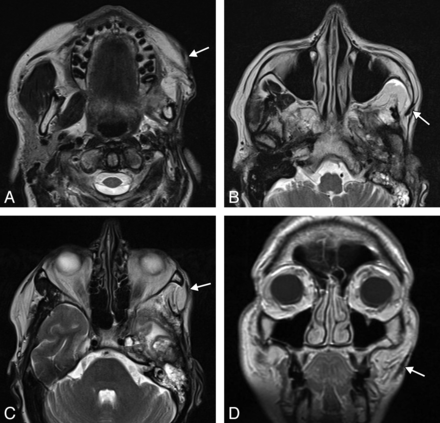
The temporalis flap (temporalis muscle transposition) procedure is an effective option for reanimation of the smile.22 An approximately 1.5-cm-wide strip of the midportion of the temporalis muscle is dissected off the calvaria and reflected in the subdermal plane from the zygomatic arch to the modiolus of the oral commissure (Fig 14). The course of the transposed temporalis muscle can be delineated on cross-sectional imaging as a thin band of soft tissue (Fig 15).
Temporalis flap. Intraoperative photographs show that the middle portion of the temporalis muscle (arrow) has been dissected free and transposed toward the oral commissure. The flap was subsequently tunneled beneath the subcutaneous tissues.
Temporalis flap. Axial T2 MR imaging (A–C) and coronal T1 MR imaging (D) show that the left temporalis muscle with the overlying fascia (arrows) is directed retrograde from the temporal fossa to the orbicularis oris.
Gore-Tex Sling Suspension
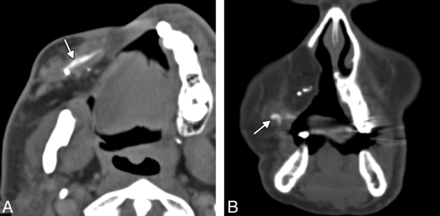
Gore-Tex (expanded polytetrafluoroethylene) (W.L. Gore and Associates, Reisterstown, Maryland) can be used for static suspension in facial paralysis.23,24 It is manufactured in thin 1- to 2-mm sheets, which can be cut into strips and implanted through small incisions. On CT, Gore-Tex slings appear as linear hyperattenuations (Fig 16).25 Although the use of Gore-Tex in facial reanimation eliminates donor site morbidity associated with the harvest of autologous grafts, the allograft is prone to complications, such as delayed wound infection.24
Gore-Tex sling. Axial (A) and coronal (B) CT images show the linear hyperattenuated strip of Gore-Tex that supports the right oral commissure (arrows). The patient is status post right complete maxillectomy with myocutaneous flap reconstruction.
Footnotes
Disclosures: Mary E. Cunnane—UNRELATED: Other: WorldCare Clinical, Comments: I was a radiologist for a clinical trial of a chemotherapy drug for head and neck cancer.
Indicates open access to non-subscribers at www.ajnr.org
REFERENCES
- © 2014 by American Journal of Neuroradiology


