Abstract
SUMMARY:Intradural extramedullary location of ependymoma is rare. To the best of our knowledge, only 9 cases have been described in the literature. We report a case of a histologically confirmed ependymoma (WHO grade II) presented in the MR imaging as a cystic, nonenhancing thoracic intradural extramedullary lesion compressing the spinal cord. The cystic appearance mimicking an arachnoid cyst at diagnosis and the leptomeningeal dissemination developed later were peculiarities that have never been previously described in relation to these rare tumors.
Tumors of the spinal cord are unusual. In a general hospital, only 5% of spinal tumors are intramedullary, 40% are intradural extramedullary, and 55% are extradural.1 Ependymomas typically have an intramedullary location and represent 60% of the intramedullary tumors.1
Primary intradural extramedullary ependymomas of the spinal cord are rare; only 9 cases have been described in the literature.2 We describe a primary extramedullary ependymoma that presented on MR imaging as an arachnoid cyst and discuss this unusual finding.
Case Report
A 67-year-old woman was admitted with a 2-year history of sensation of “sleeping feet” and “tight socks.” The most relevant complaint was gait impairment and instability due to loss of deep tendon sensation. Neurologic examination showed disturbed sense of position with preserved tactile and temperature sense, normal muscular force, and normal reflexes. Electrophysiologic evaluation excluded a sensitive polyneuropathy.
Spinal MR imaging was performed by using a spine surface coil and a 44-cm field of view. Sagittal spin-echo T1-weighted (TR, 400 milliseconds; TE, 12 milliseconds) and T2-weighted (TR, 2473.4 milliseconds; TE, 110 milliseconds) images were acquired. Two axial T2-weighted sequences (T2 gradient-echo: TR, 594.8 milliseconds; TE, 13.8 milliseconds; T2 driven equilibrium radio frequency reset pulse: TR, 1000 milliseconds; TE, 1000 milliseconds) were also obtained. After gadolinium injection, axial and sagittal enhanced T1-weighted images (TR, 400 milliseconds; TE, 12 milliseconds) were acquired.
The images (Fig 1) demonstrated an extramedullary lesion, posteriorly located and extending from T5–T6 to T8, compressing the spinal cord anteriorly. The axial images (Fig 2) were the most useful in demonstrating the extramedullary location. The lesion was well defined, with some septa inside. It was essentially isointense with the CSF on all pulse sequences. The adjacent spinal cord was abnormal on T2-weighted sequences with a hyperintense appearance (T5–T6) (Fig 1A,-B). After gadolinium injection (Fig 1C,-D), there was no enhancement of the cystic extramedullary lesion. No enhancement was seen within the spinal cord, but the cord surface was covered by a rich vascular, enhancing network, probably because of the compression of the venous plexus system. No other abnormalities were found within the brain or in the spinal cord. The final preoperative diagnosis was arachnoid cyst with spinal cord compression.
Preoperative MR imaging. Sagittal T2- (A and B) and T1-weighted images before (C) and after (D) gadolinium injection. It is extremely difficult to determine whether this lesion is intra- or extramedullary. No contrast enhancement is seen. Septa are observed within the lesion.
Axial T2-weighted images at different thoracic levels (A and B) better illustrate the extramedullary location of the lesion (arrow) and its posterior, lateral right position.
A bilateral dorsal T6–T7 laminectomy was performed, and, following the opening of the dura mater, a cystic lesion with well-limited thick walls containing multiple noncommunicating pockets of arachnoid membranes was found. The lesion was purely extramedullary (Fig 3). There was no attachment of the lesion to the spinal cord or to the dura mater, but an important arachnoid inflammatory reaction was visualized next to the lesion. Complete resection of those membranes was performed.
Macroscopic appearance of the lesion at surgery after opening of the dura mater: a cystic mass mimicking an arachnoid cyst, with abnormally thick walls.
Histologic examination (Fig 4) of the surgical specimen showed thickened arachnoid related to tumoral proliferation constituted of monomorphic cells with regular round nucleus and dispersed chromatin. Tumor cells of the perivascular area were arranged radially around the blood vessels (Fig 4C) forming perivascular pseudorosettes with prominent immunoreaction for glial fibrillary acid protein (GFAP) (Fig 4D). No true ependymal rosettes were observed. Mitoses and necrosis were absent. Final histologic diagnosis was a WHO grade II ependymoma.
Histologic examination reveals an ependymoma grade II. A, Low-power view illustrates tumor proliferation located around the arachnoid. B, Thickened arachnoid is limited but not invaded by monomorphous tumor cells. The cyst wall is shown at the periphery of the specimen. C, Perivascular cellular arrangement around hyalinized blood vessels denotes ependymoma differentiation. D, GFAP immunostaining confirms the presence of pseudorosettes.
Regression of the symptoms, primarily the gait impairment, occurred after surgery. The immediate postoperative MR imaging (Fig 5) showed total resection of the retromedullary cyst. Surprisingly, a new intradural extramedullary cyst was found at the level of T6, anteriorly located but still compressing the spinal cord. This new cyst showed the same characteristics of the previous one. There was persistent abnormal signal intensity within the spinal cord on T2-weighted images (Fig 5A).
Postoperative MR imaging. Sagittal T2- (A) and T1-weighted images before (B) and after (C) gadolinium injection; axial T1- (D) and T2-weighted (E) images. A ventral intradural extramedullary T6 lesion is found, presenting the same features as the previously removed lesion.
Progressive worsening of the gait impairment and sensitive deficits were noted, and surgery was performed again 10 months after the first surgery. Perimedullary tumor not attached to the spinal cord was again found. Histologic examination confirmed an ependymoma, WHO grade II. The immediate postoperative MR imaging (Fig 6) showed complete macroscopic removal of the lesion.
Sagittal T2-weighted (A) and T1-weighted gadolinium-enhanced (B) images obtained after a second surgery showing the total removal of the anteriorly located cystic lesion. Persistent spinal cord hypersignal intensity is seen in T2-weighted image (arrow).
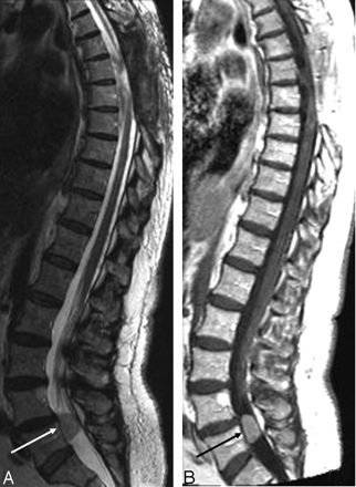
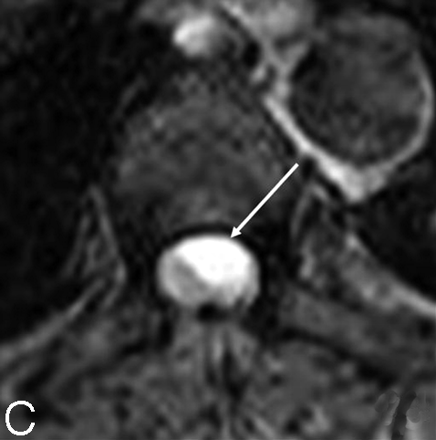
Three months after this second operation a control MR imaging (Fig 7) demonstrated, at the site of prior surgery, a recurrent cystic lesion behaving in a manner similar to that observed on the previous studies and also a new intradural extramedullary lesion at the level of L5. A homogeneous nodule was found with a slightly hyperintense aspect on T2-weighted-images (Fig 7A) enhancing strongly after contrast administration (Fig 7B). This new lesion was considered to be a drop metastasis, and surgery again confirmed an ependymoma grade II, though the behavior of the tumor was clinically aggressive.
Control MR imaging examination. Sagittal T2 (A) and T1-weighted gadolinium-enhanced (B) images disclosing an intradural L5 drop metastasis (confirmed by subsequent surgery). Moreover, at the site of prior surgery, the cord looks again newly deformed by a recurrent cystic lesion behaving in a similar way observed on the previous studies. This aspect is confirmed on the axial T2WI obtained at the T7 level (C): the cyst is left anterolaterally located (arrow).
Discussion
Primary intradural extramedullary ependymomas of the spinal cord are extremely rare, in contrast to intramedullary ependymomas or ependymomas arising from the conus medullaris or filum terminale.2-5 Occasional cases of ectopic ependymoma outside the spine have been mentioned, developing in the subcutaneous tissue over the sacrococcygeal region, the meso-ovarium, the ovary, and the broad ligament.2,6 The origin of intradural, extramedullary ependymomas is uncertain. They probably arise from heterotopic glial tissue pinched off from the neural tube during its closure.2,6,7 In total, 9 cases are described in the literature. They mimic the clinical presentation and the radiographic characteristics of more common intradural extramedullary lesions such as neurinomas, meningiomas, or, rarely, as in our case, an arachnoid cyst.
All cases reported in the literature concern women: the hypothesis of a hormonal mechanism playing a role in the development of the tumor was suggested by Duffau et al.2
A history of progressive medullary compression was usually reported.2 In our patient, the symptoms were related to the posterior extramedullary location of the lesion.
In the literature, a thoracic location is found in 7 cases. One cervical location,5 and an ependymoma arising from the L3 nerve root6 were described as well.
None of the described cases had similar imaging features compared with our patient: especially the cystic appearance mimicking an arachnoid cyst has never been reported. The rare extramedullary ependymomas described in the literature were solid tumors showing a hyper signal intensity on long TR/long TE images and isosignal intensity on short-TR/short-TE images. Moreover, those tumors exhibited a moderate to intense enhancement, very similar to meningiomas or neurinomas.2
In the present case, the intraoperative findings were consistent with multiple, isolated cystic lesions, without any attachment to the central nervous system or to the dura mater. The lack of connection between the lesion and the central nervous system excludes the hypothesis of an exophytic ependymoma of the spinal cord.8,9 An ependymal metastasis is equally unlikely due to the absence of any primary cerebral or medullary tumor.
In our case, apparently new anteriorly located lesions have developed at the same level and with the same characteristics, 1 month after the first surgery and 3 months after the second surgery. In our opinion, these lesions are more probably recurrent than residual lesions, originating from a microscopic residual ependymoma, though early recurrence is not described in the cases reported.2 Another explanation is that the residual lesions were hidden by compression of the spinal cord. Surgical decompression permitted re-expansion of these predominantly cystic anomalies.
Another peculiarity of our case was the occurrence of an intradural drop metastasis 14 months after the diagnosis of a primary, cystic tumor had been made. In the literature, no meningeal dissemination of an intradural extramedullary ependymoma has been reported. Complete resection, if technically possible, and postoperative local radiation therapy for incomplete resection in case of malignant intramedullary ependymoma are recommended.10 We can thus assume that the same treatment strategy is also recommended for extramedullary ependymoma.
In conclusion, extramedullary ependymoma is a rare diagnosis that may mimic other lesions and should thus be considered in the differential diagnosis of intradural extramedullary lesions.
- Received May 6, 2005.
- Accepted after revision June 3, 2005.
- Copyright © American Society of Neuroradiology


