
Abstract
BACKGROUND AND PURPOSE: The purpose of this study was to evaluate the presence of blood clots in femoral arterial sheaths maintained after cerebral angiography and the effect of heparinized saline on clot formation.
METHODS: Twenty-three sheaths were evaluated in 18 patients. Sheaths were maintained for 14 to 80 hours (average, 33 hours; median, 24 hours). After the sheaths were removed, they were vigorously flushed with 60 mL of normal saline and the number and size of clots found in each sheath were recorded. Additionally, patients' age, catheter size, presence of heparin, amount of time the sheath was kept in the artery, and patients' coagulation status were recorded.
RESULTS: Clots were found in 17 (74%) of the 23 sheaths. Ten catheters had continuous heparin drip, of which seven (70%) sustained clots. Of the 13 sheaths without heparin, 10 sustained clots (77%). The difference was not statistically significant. The average number of clots was 2.2, and the maximal length of clots ranged from 0.5 to 105 mm. No thromboembolic complications associated with sheath placement were encountered in our patient population.
CONCLUSION: Blood clots are present in the vast majority of intraarterial sheaths maintained after cerebral angiography. These clots constitute a risk of thromboembolic complications in the event of repeat angiography. Sheath exchange should be considered before obtaining repeat cerebral angiograms.
The use of femoral sheaths for arterial vascular access is a common interventional neuroradiologic practice. Sheaths are used for both short-term access in routine diagnostic angiography as well as for prolonged neurointerventional procedures. Typically, diagnostic angiographic procedures use smaller access sheaths of 4F to 6F, whereas interventional neuroradiologic procedures frequently require larger guidecatheters, necessitating the placement of larger femoral sheaths. The latter are usually in the 6F to 10F range, depending on the needs of a specific case.
Commonly, a heparinized saline flush is used in an effort to prevent in vivo clot formation within the sheath lumen. However, the use of heparin in arterial sheath flush lines is controversial, because it may aggravate acute intracranial hemorrhage. With the introduction of percutaneous treatment techniques for cerebrovascular disorders, particularly the use of detachable coils in the treatment of ruptured aneurysms, the short-term use of heparin has became common practice in reducing thromboembolic catheter-related complications. However, the relatively prolonged use of heparin in flush lines remains controversial, regardless of whether the patient requires percutaneous or surgical intervention.
Indications for long-term arterial sheath placement include the need for follow-up angiograms after clipping or coiling of intracranial aneurysms, the anticipated use of intraoperative angiography in patients with previous arterial sheath placement, and the need for numerous repeat follow-up angiograms for intracranial vasospasm treatments, arterial blood pressure monitoring, or simply until systemic anticoagulation is reversed, allowing for safe sheath removal. This study was undertaken to evaluate the effect of heparinized flush versus saline flush on clot formation within arterial sheaths maintained overnight.
Methods
Patients were included in our study if a femoral arterial sheath was maintained for more than 12 hours. Sheaths included in this study were placed and removed by our neuroradiology service. All sheaths were manufactured by Terumo (Tokyo, Japan) under the brand name Pinnacle. Patients were randomized into two groups, one receiving heparinized saline (2000 U/L) and the other receiving normal saline. All sheaths were maintained with a transducer-monitored flushing system, with a flush rate of 3 mL per hour, according to our intensive care unit protocol. Each time a study sheath was removed from a patient, the sheath was immediately evaluated at bedside for indwelling clots by means of vigorous flushing with 60 mL of normal saline solution. The sheath was then examined for the presence of any residual clots. The number and size of clots were recorded. Additionally, the patients' age and sex, the catheter size, the duration of sheath placement, and the patients' coagulation status during the time the sheath was in place were recorded (see Table).
Patient characteristics
Results
Twenty-three sheaths were evaluated in 18 patients (see Table). Sheaths were maintained for 14 to 80 hours (average, 33 hours; median, 24 hours). Clots were found in 17 (74%) of the 23 sheaths. Ten catheters had continuous heparin drip, of which seven (70%) sustained clots. Of the 13 sheaths without heparin, 10 sustained clots (77%). The difference was not statistically significant (P = 1.0, Fisher exact test). The average number of clots was 2.2, and the maximal length of clots ranged from 0.5 to 105 mm. No thromboembolic complications associated with sheath placement were encountered in our patient population. One of the larger clots obtained in our study group is shown in Figure 1.
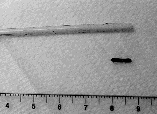
Photograph of a typical clot obtained from an arterial sheath
Discussion
Known complications of femoral arterial puncture include groin hematomas, pseudoaneurysm formation, local femoral or iliac arterial dissection caused by local arterial wall trauma, infection, retroperitoneal hemorrhage, arteriovenous fistula, and emboli, with an overall complication rate of 0.5%. The risk of thromboembolic complications related to prolonged maintenance of femoral arterial sheaths is low (1–5).
Findings reported in the current nursing literature support the use of saline flushing alone in the maintenance of peripheral venous access or arterial pressure monitoring catheters without added heparin because of its proved efficacy in maintaining the patency of both peripheral intravenous locks and radial arterial pressure monitoring lines (6–9). Interventional neuroradiology practices, however, are unique, in that typically larger caliber sheaths are used and may be repeatedly traversed with inner catheters for follow-up angiograms. Clots were found in 77% of the nonheparinized sheaths and in 70% of the heparinized sheaths. We found the difference to be not statistically significant, possibly because of the small number of patients in our series. However, even if heparin does reduce the risk of clot formation in the sheath, it definitely does not prevent clots from forming.
Before this study, we did not exchange an indwelling sheath before every repeat neuroangiographic study. It has not been our experience to see distal embolic complications when reusing sheaths that had not been exchanged. However, we theorize that there is potential, though small, for small emboli to be pushed out of the sheath into the arterial tree by an inner catheter. We suspect that these emboli are frequently silent and most likely lyse without sequelae. However, larger clots, as large as the ones seen in our study, may be a problematic cause for significant thromboembolic complications.
Conclusion
This study confirms that small emboli do form within indwelling femoral arterial sheaths maintained “overnight.” This occurs regardless of the addition of heparin to the flush solution. In this study, we did not evaluate the effect of flush rates on clot formation. We can only speculate that, when possible, a faster rate of flushing can add an additional level of effectiveness in the prevention of clots within sheaths. Therefore, we advise caution when reusing long-term arterial sheaths for follow-up angiographic procedures. In the event of repeat cerebral angiographic procedures through an existing femoral arterial sheath, sheath exchange should be strongly considered.
Footnotes
↵1 Address reprint requests to Robert A. Koenigsberg, DO.
- Received June 22, 1998.
- Copyright © American Society of Neuroradiology


